The formation of pulp stones has also been. Pulp stones are discrete calcifications that form within the pulp chamber.
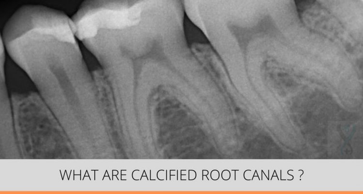
What Are Calcified Or Blocked Root Canals Expert Dental Care
It is believed that the.

. They may lie free within the pulp adhere to the chamber wall or become embedded in dentin. The dental status was scored as. There are several reasons that a pulp chamber will have calcification.
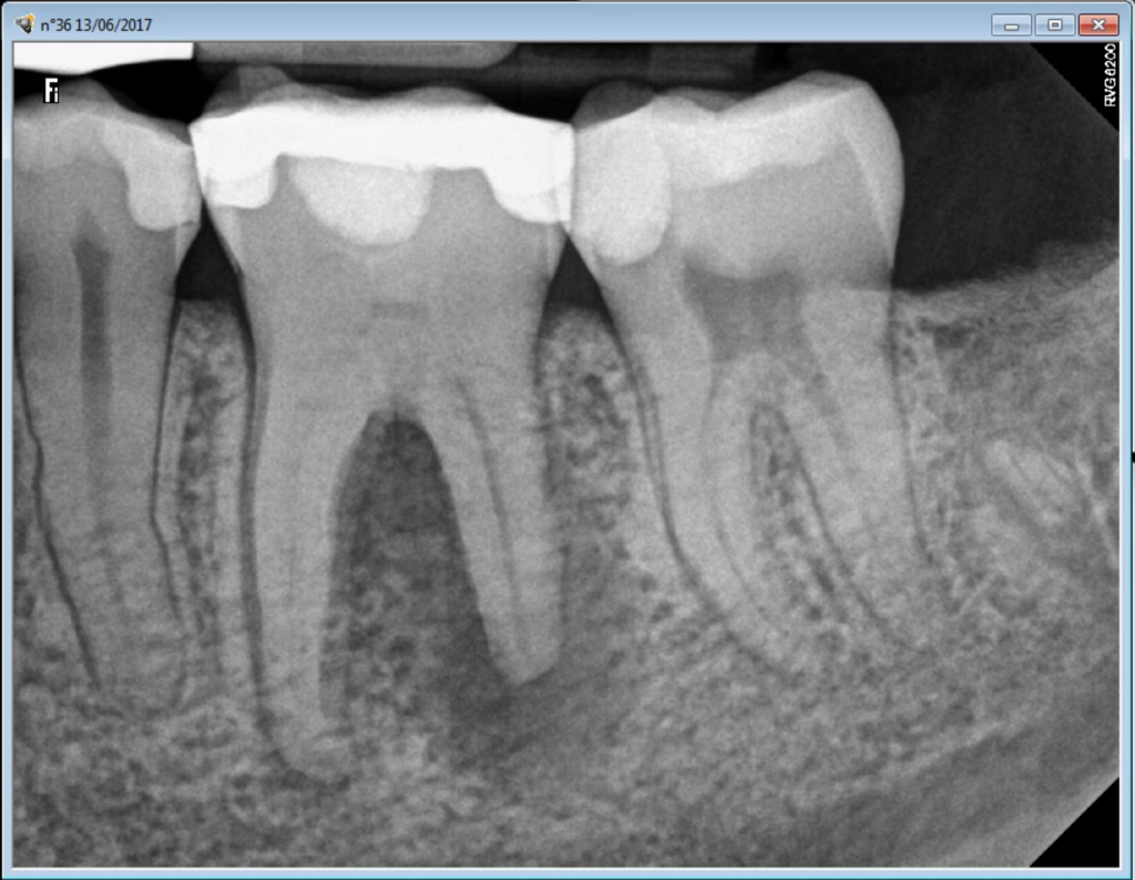
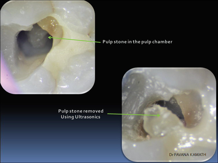
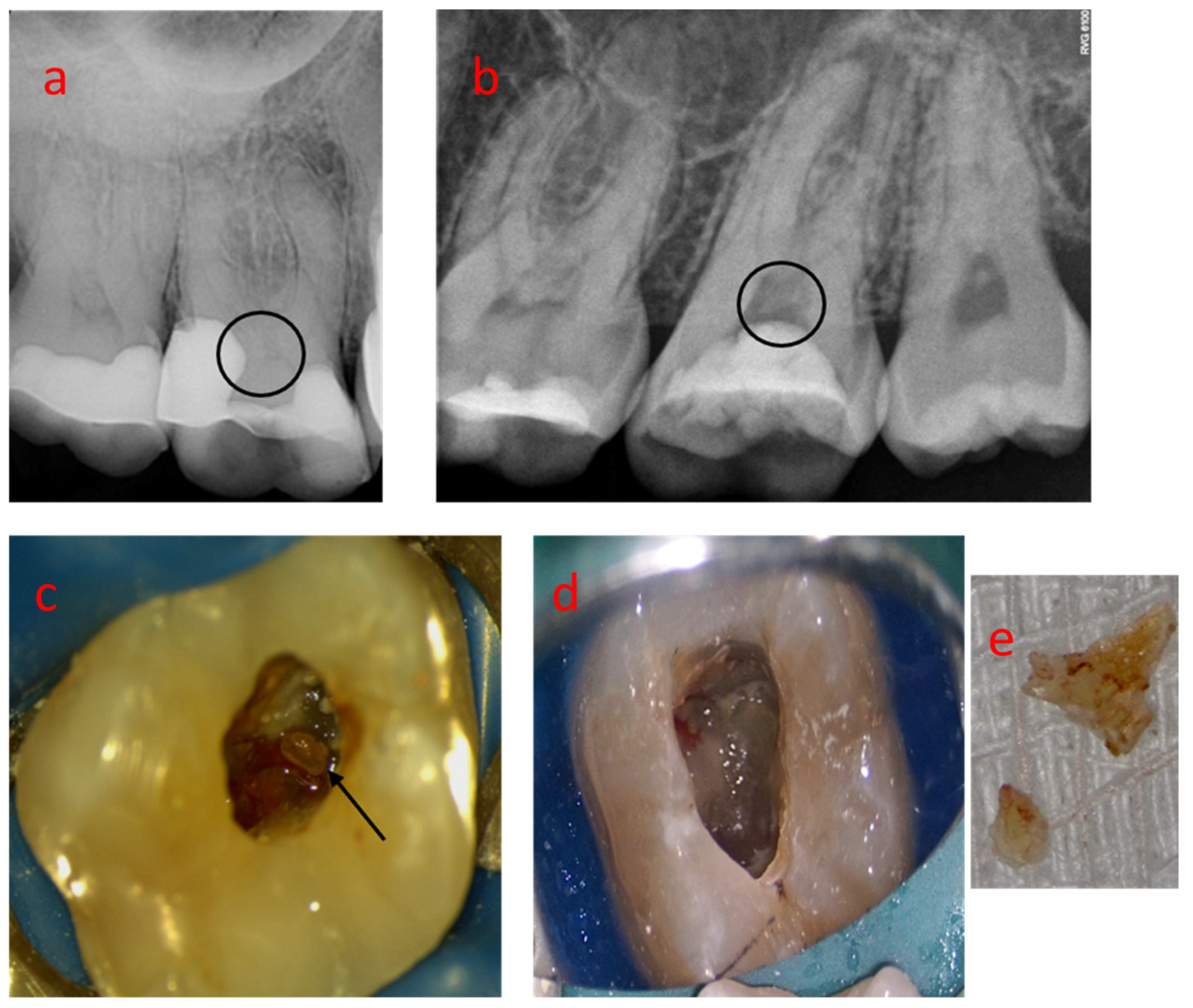
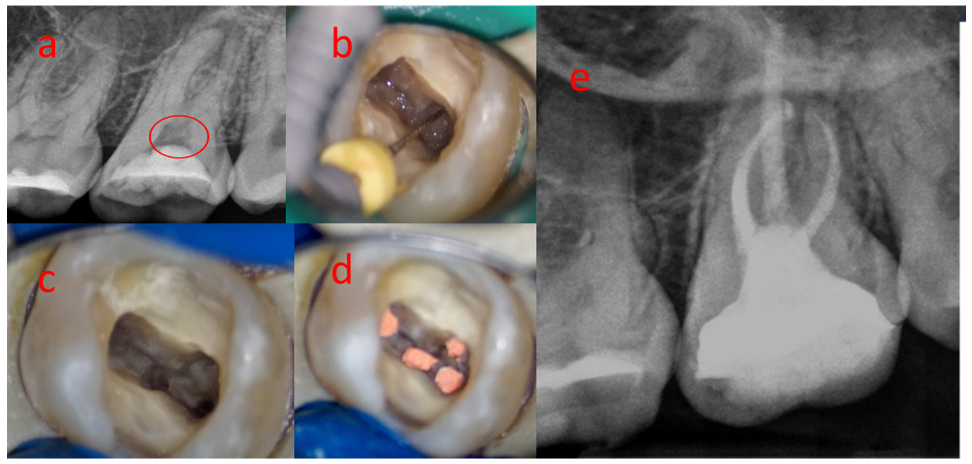
A Magnified view of the pulp chamber after removal of the amalgam and secondary car-ies. Gaining access through a calcified pulp chamber. Pulp calcifications stones are nodular calcified masses appearing in either or both the coronal or root portions of the pulp organ.
A clinical challenge Abstract Dental pulp is prone to dystrophic mineralization. The pulp calcifications were classified as diffuse or discrete and their locations were recorded as root or crown. The recommended clinical steps for our modified technique include.
After achieving access to the pulp chamber using a high speed turbine place the ultrasonic tip in direct contact with the pulp calcification. Pulp stones are primarily a physiological manifestation as are most other pulpal calcifications and may increase in number andor size. A brief description of management of calcified pulp chamber and canals is given here.
When a dental professional takes an X-ray of a tooth with calcification they may notice its abnormal appearance. The role of a calcium. The term calcified tooth means a tooths normal dental pulp chamber is compromised due to a reduction in size or obliterated due to trauma disease decay or age.
The root canal space. The dental pulp calcification presents as masses of calcified tissue present on the level of the pulp chamber and roots of the teeth15. Since those are common and known to all dentists I will not touch on those causes.
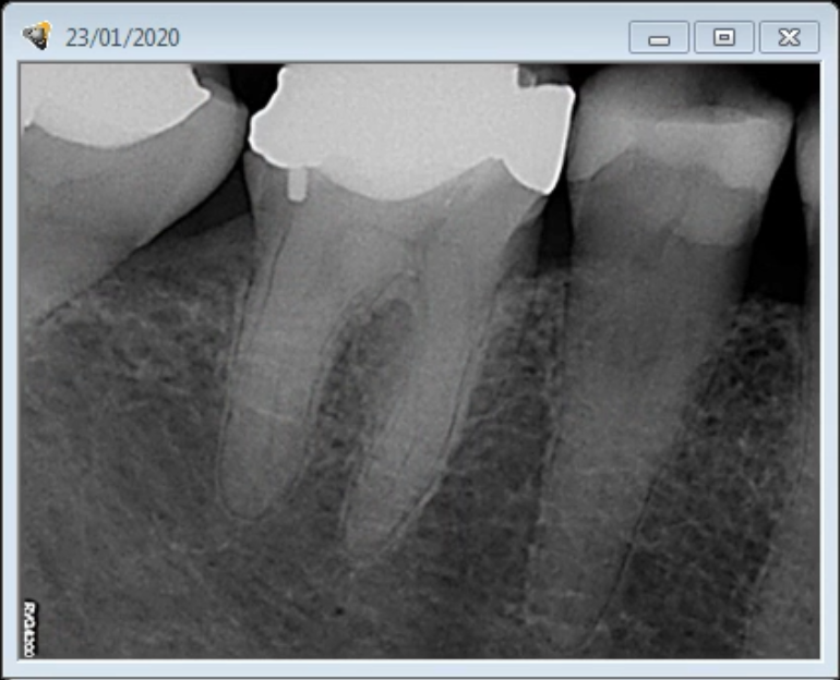
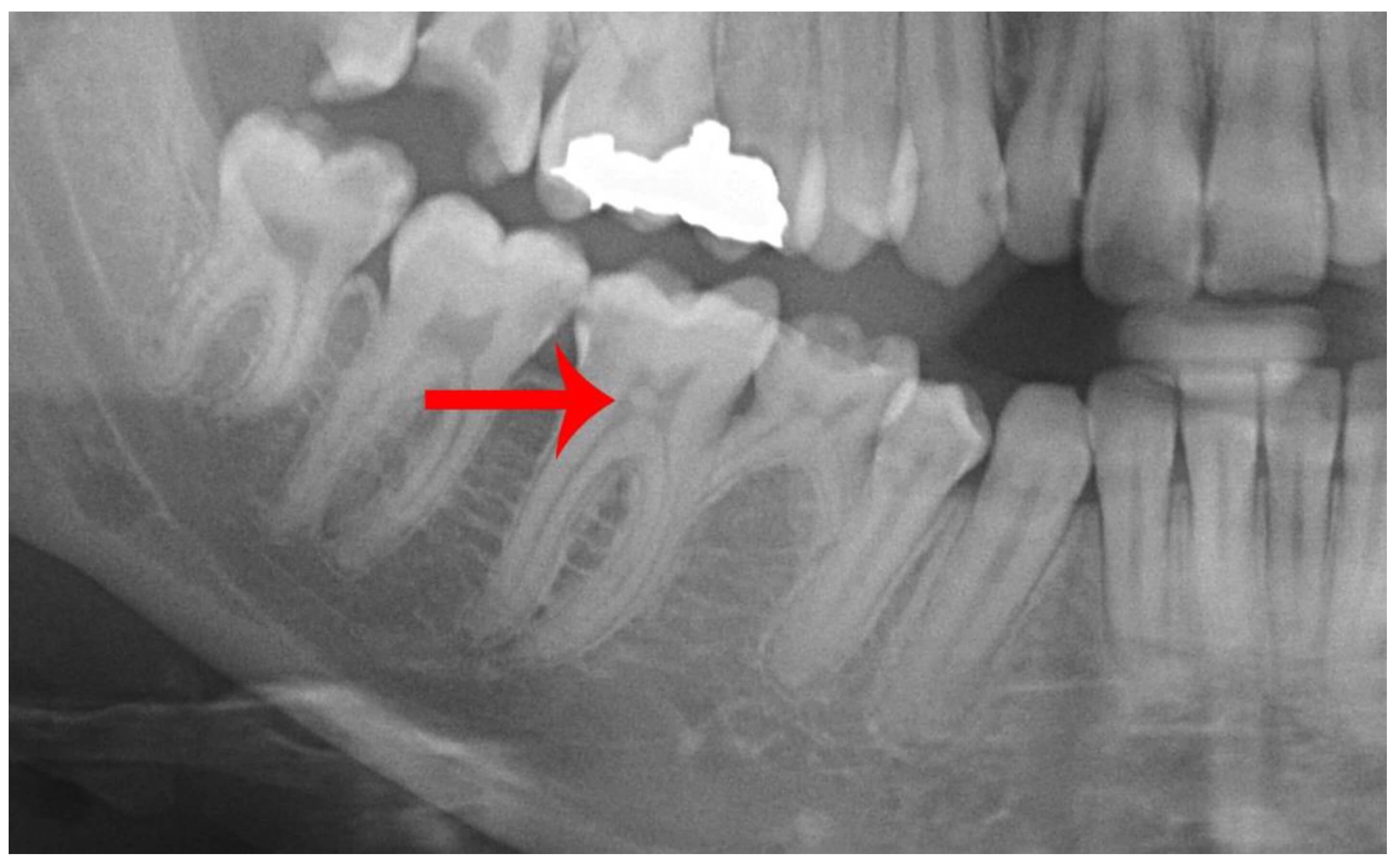
Note the evidence of a. Pulp chamber is almost completely obliterated by adherent calcifications. When the pulp chamber was completely radiolucent that tooth was scored as tooth without pulp chamber calcification.
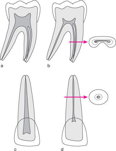
The height of a pulp chamber is between 15 to 20 mm Figure 5a. The other causes are. This article presents the endodontic management of a tooth with an obliterated pulp chamber and associated with a discharging sinus in a teenage patient.
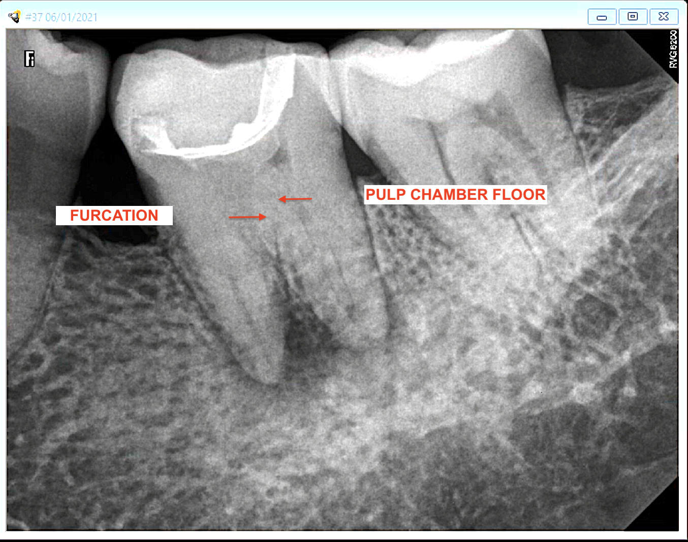
ACCESS CAVITY PREPARATION After initial clinical and radiographic examination the quality of the coronal restoration if present should be checked and insufficient restorations. Step-by-Step Calcified Canal Location 1. Of the pulp chamber and partial calcification of the root canals.
This 15-mm to 20-mm measurement is the most variable due to calcifications because of aging caries and. Root canal entries are also embedded. As an IJHS review explains the X-ray may show that the pulp chamber.
Calcific metamorphosis or pulp canal obliteration PCO is the pulp response to trauma characterized by rapid deposition of mineralized tissue in the root canal space. When the pulp chamber was completely radiolucent that tooth. Definite radiopaque focuses inside the radiolucent pulp chamber were defined as pulp chamber calcifications.
2Sealing the floor of the access cavity with. They often develop in teeth that appear quite normal in other. 1Access cavity preparation in the pulp chamber without entering the canal.
As pulp chamber calcifications. The role of a calcium hydroxide lining to induce mineralization and cause the obliteration of the pulpal space is also discussed. The most common that dentists see everyday all day is local trauma or natural aging.
Root Canal - Calcified Pulp Chamber With a Guide. The discrete calcifications were further classified according to. Microscope could not be of some use at the beginning of.
The maxillary molar endodontic access opening. When the first most coronal pulp chamber floor has been reached with a 330 long shank bur the root canal entries were still obscured by reparative dentine and could not be. This article presents the endodontic management of a tooth with an obliterated pulp chamber and associated with a discharging sinus in a teenage patient.
This mineralization can be so extensive that the entire root.

Endodontic Management Of A Tooth With Large Pulp Stone Style Italiano Endodontics
Pulp Obliteration And Root Canal Endomontreal

What Is A Calcified Root Canal And How To Locate It Root Canal Treatment
Saving A Severely Calcified Tooth With A Root Canal Procedure Endomontreal
Management Of Calcified Pulp Chamber Canals With Ultrasonics Youtube
Coronally Cut Sample Exhibiting Calcified Pulp Stone Arrow And Download Scientific Diagram
Root Canal Calcifications A Challenge In Root Canal Treatments Endomontreal

A Preoperative Radiograph Showing Diffused Radio Opacities Throughout Download Scientific Diagram
What Are Pulp Stones News Dentagama
Dental Pulp Calcification And Changes With Age Bite Point
Role Of Ultrasonics Tips Under Magnification In Endodontics 1
Dental Pulp Calcification And Changes With Age Bite Point
Case 4 Calcified Right Lower First Molar A Preoperative Radiograph Download Scientific Diagram

Dental Pulp Calcification All The Causes Bauer Smiles
5 Entering Calcified Systems Pocket Dentistry
2

Endodontic Management Of A Tooth With Large Pulp Stone Style Italiano Endodontics
Medicina Free Full Text The Pulp Stones Morphological Analysis In Scanning Electron Microscopy And Spectroscopic Chemical Quantification Html
Medicina Free Full Text The Pulp Stones Morphological Analysis In Scanning Electron Microscopy And Spectroscopic Chemical Quantification Html
